Laparoscopy & Endoscopy
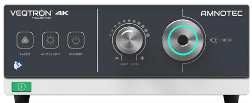
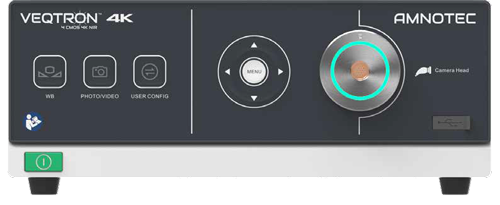
VEQTRON® ICG/NIR Twinlight LED-LASER
LED and laser light source for standard white light as well as ICG/NIR light mode.
View details
Laparoscopy & Endoscopy
Brand: VEQTRON®
ICG/NIR fluorescence imaging for minimally invasive surgery, with 4 CMOS 4K technology and four display modes.
ICG/NIR fluorescence imaging has become one of the most valuable technologies in minimally invasive surgery, especially for intra-operative checks of visceral perfusion, the visualisation of biliary structures and lymph node mapping.
The VEQTRON Imaging System offers four different display modes without any loss of image quality.
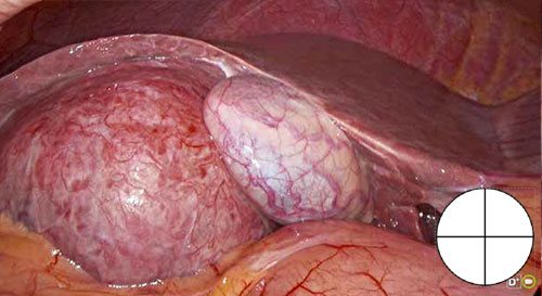
3-CMOS white light image, used for all standard minimally invasive surgeries.
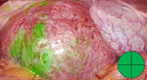
High-quality fluorescence image for detection of ICG.
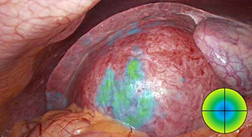
Fluorescence and white light image merging for intensity tracking of ICG direction of flow.
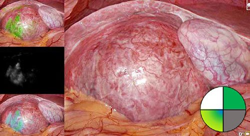
White light, fluorescence green, ISO fluorescence and NIR black-and-white displayed at the same time, with the option to choose the mode on the main display.
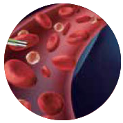
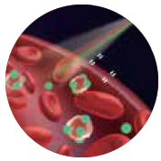
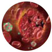
Fluorescence refers to the ability of some atoms and molecules to absorb light at a particular wavelength (excitation) and then to emit light of a longer wavelength during a short-lived emission. This means:
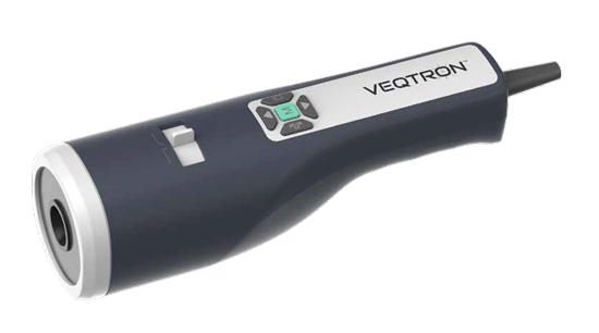
As of today, we are still at the beginning of the possibilities for indications with fluorescence. Due to new innovative techniques, such as the hand-held camera, fluorescence will also become more and more important in open oncological surgical techniques, e.g. in the parathyroid gland (autofluorescence).
Laparoscopy & Endoscopy
LED and laser light source for standard white light as well as ICG/NIR light mode.
View detailsLaparoscopy & Endoscopy
Handheld fluorescence camera for open surgery.
View detailsLaparoscopy & Endoscopy
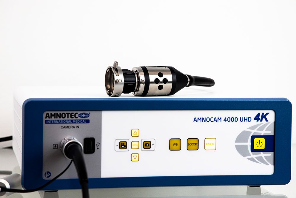
Camera and video devices using 4K Ultra HD, HD-MOS and PHASER™ technology.
View detailsLaparoscopy & Endoscopy
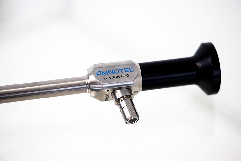
High-resolution endoscopes in stainless steel with scratch-resistant sapphire glass, autoclavable and approved for…
View details